
Nuclear Medicine
About the Department
The Nuclear Medicine Department at the Warith International Cancer Institute offers various services, primarily focused on cancer patients. One of the most important tests provided by nuclear medicine is the PET/CT scan (Positron Emission Tomography/Computed Tomography) for cancer patients, which is a cornerstone in tumor diagnosis and determining the tumor stage, as well as assessing the patient's response to chemotherapy, radiation therapy, and hormone therapy.The Bone Scan is a nuclear imaging test that determines whether there is bone metastasis. There are other tests and services provided by nuclear medicine for patients not diagnosed with tumors, including kidney and urinary tract tests such as DTPA, MAG3, and DMSA, as well as nuclear imaging of the thyroid (Thyroid Scan) and the parathyroid glands (Sestamibi scan).
The MIBG scan is precisely used to diagnose adrenal gland diseases and neuroendocrine disorders, as well as to determine their stage and response to treatment. The Nuclear Medicine Department at the Warith Institute is the only center in Iraq that offers this scan to patients from north to south.
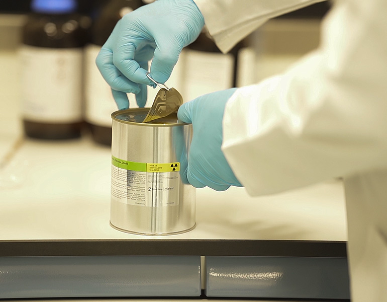
In addition to diagnostic services, the Warith Center for Radiopharmaceutical Therapy offers treatment for benign and malignant thyroid tumors, with doses ranging from 10 millicuries to 250 millicuries.
The Nuclear Medicine Department is a branch of medicine that includes both diagnostic and therapeutic components. It uses small amounts of radioactive materials to diagnose, assess the severity, and treat various diseases, including many types of cancers, heart conditions, digestive disorders, glandular disorders, neurological conditions, assess kidney functions, distinguishing obstructions in the urethra and urinary tract, and other bodily changes. As nuclear medicine procedures can pinpoint precise activities within the body, they offer the ability to detect diseases at early stages and evaluate the response to medical interventions.
The Diagnostic Department:
Nuclear medicine scans are painless tests that help doctors diagnose and assess medical issues. These scans use radioactive materials called radiopharmaceuticals or radiotracers, administered via intravenous injection or orally, depending on the type of nuclear scan. The emitted radiation is then captured by a special camera or imaging device that generates images and provides precise information.
The specialist doctor sees patients in the consultation area, whether they are inpatients or outpatients (usually patients with thyroid conditions, both benign and malignant). The consultant evaluates patients with metastatic prostate cancer resistant to other treatments to coordinate and assess their suitability for targeted radiopharmaceutical therapy, including PSMA LU177. Additionally, patients with neuroendocrine tumors resistant to other treatments can be assessed for potential treatment with LU177 DOTATATE.
The specialist doctor performs a clinical examination, conducts necessary tests, and takes required imaging studies to assess the extent of the disease and determine the appropriate doses of radioactive iodine (1131) to be administered.
Patients are given special instructions on the iodine diet to be followed two weeks before the dose date as well as post-biopsy instructions for therapeutic radioactive iodine at doses greater than 30 mCi.
Patients treated with radioactive iodine at doses above 30 mCi are admitted to the Warith International Cancer Institute (WICI) for Radiopharmaceutical Therapy for one or two days, as measured by the residual radiation in the patient's body by a specialized dosimeter.
We inform the patient about the nuclear scan that needs to be performed in the gamma camera department one week after administering the therapeutic capsule and two days after administering the diagnostic capsule.
The specialist doctor writes the report, informs the patient about the treatment results, and monitors the condition every 6 months.
The most important tests:
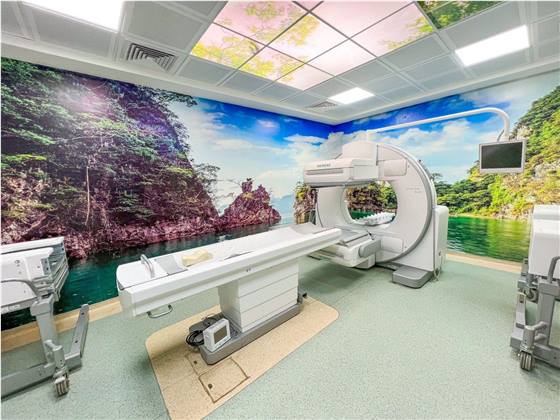
• Gamma Camera: Gamma camera device.• PET/CT scan: Positron emission tomography/computed tomography scanner.
• RAI: Radioactive iodine.
Cyclotron
It is the primary link and key factor in producing radiopharmaceuticals used in diagnosing tumors, heart diseases, and neurology, and is one of the most important components of the department. It is a European-manufactured (Belgian) cyclotron, which produces fluorodeoxyglucose (FDG) through several complex steps using heavy oxygen-18 as the starting material. The device also features a very high production capacity, enabling it to supply most centers in Iraq with this important substance used in PET scans for accurate cancer cell detection. Additionally, the device has the capability for future development to produce various radioactive materials such as Gallium-68, Carbon-11, and Nitrogen-13, and others. It is designed to accommodate future expansions and has eight production ports that can be used to add production lines as needed. The current production capacity of the cyclotron at the Warith Institute is 10 curies per cycle.What is a cyclotron?
A cyclotron is a particle accelerator that uses electromagnetic fields to propel particles to produce radioactive isotopes needed for positron emission tomography (PET) for various applications, including oncology.
Importance:
It is the primary link and key factor in producing radiopharmaceuticals for diagnosing tumors, heart diseases, and neurology.
Features:
It is distinguished by its very high radioactive production capacity, allowing it to supply most PET scan centers in Iraq with fluorodeoxyglucose (FDG) while adhering to the highest standards of quality control and quality management, and it has the potential for future development to produce other radioactive isotopes.
Cyclotron mechanism:
The device is the largest in Iraq, with a nuclear coupling capacity that can be incrementally increased to 100 MA and an energy level of 18 MeV, reaching up to 300 MA. It can produce radioactive doses of FDG sufficient for scanning 60 to 100 patients in a single operating cycle of 120 minutes. Additionally, the device features 8 ports, providing high flexibility for producing a variety of radioactive isotopes.
GAMMA Camera scans
Kidney and urinary tract imaging
Instructions:
• You may eat normally before your appointment.
• Please arrive 30-45 minutes before your scheduled time.
• You should increase your fluid intake, such as water, juice, and tea.
• The total amount of fluids consumed is 1.5-2 liters before the exam.
• The exam does not require you to stop any medications you are currently taking.
• You will need to wait for 2-4 hours after receiving the injection.
• The imaging procedure will take between 15-30 minutes.
• Empty your bladder before the scan.
PET/CT scan
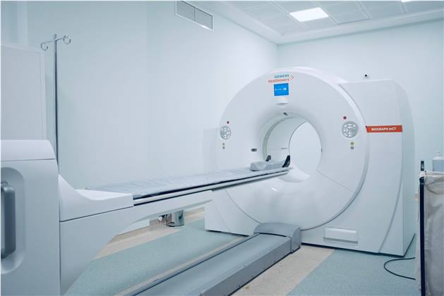
The PET/CT scan is a non-invasive, painless procedure that takes approximately 20-30 minutes. In addition to providing better imaging data, it enhances patient comfort by reducing the number of scans needed. Positron emission tomography/computed tomography (PET/CT) imaging enables more precise diagnoses and developments, allows for more targeted treatment plans, and provides less invasive monitoring. This results in improved patient outcomes by accurately assessing treatment effectiveness and patient response.
Common Uses of PET/CT:
• Identifying and diagnosing cancer.
• Determining if cancer has spread throughout the body.
• Evaluating the effectiveness of treatment.
• Checking if cancer has returned after treatment.
• Assessing diagnoses.
• Evaluating tissue metabolism and viability.
• Identifying the effects of myocardial infarction or heart attack on heart regions.
• Determining which areas of the heart might benefit from angioplasty or coronary artery bypass surgery (in conjunction with myocardial perfusion imaging).
• Assessing brain abnormalities, such as tumors, memory disorders, seizures, and other central nervous system disorders.
• Mapping normal brain and heart functions.
Examinations:
1-18F-FDG PET\CT.
2-68Ga PSMA For prostate cancer.
3-68Ga DOTATATE for Neuroendocrine tumor.
4-68Ga FAPI for others.
| Gamma camera SPECT/CT | PET/CT images |
| -Tc99m MDP bone scan | -F18 FDG PET/CT for oncology |
| -Renal studies (MAG-3, DTPA & DMSA) | -Ga68 PSMA for prostate cancer |
| -Tc99m pertechnetate thyroid scan | -Ga68 DOTATATE for Neuroendocrine |
| -Tc99m sestamibi parathyroid study | -tumor, well differentiated NB & pheochromocytoma |
| -I131 MIBG study | -Tc99m nanocolloid lymphoscintigraphy |
| -Tc99m nanocolloid sentinel lymph node- I131 diagnostic whole body scan |
Available Treatments in the Nuclear Medicine Department
| I131 (10-29 mCi) doses for benign thyroid diseases include: Grave’s disease, toxic MNG, and toxic nodule |
|
| Therapeutic doses for thyroid cancer are as follows: | |
| -Targeted therapy for mCRPC I131 (30 mCi) dose |
- Lu177 PSMA |
| I131 (75 mCi) dose | I131 (50 mCi) dose |
| I131 (100 mCi) dose | Targeted therapy for metastatic Neuroendocrine tumor |
| I131 (150 mCi) dose | - Lu177 DOTATATE |
| I131 (200 mCi) dose | |